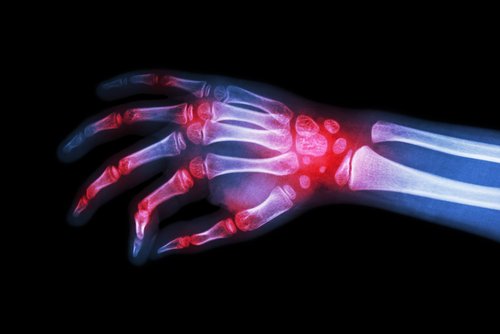
Children with recent-onset juvenile idiopathic arthritis (JIA) did not show signs of wrist damage over two years of treatment targeting inactive disease, according to a clinical trial in the Netherlands.
The findings also showed improved bone mineral density (BMD) in children with the lowest values at the start of the study, who then were treated with methotrexate and Enbrel (etanercept, by Amgen).
The study, “No radiographic wrist damage after treatment to target in recent-onset juvenile idiopathic arthritis,” was published in the journal Pediatric Rheumatology.
Bone deformity and loss, as well as cartilage loss in certain regions of the wrist, can be complications of inflammation in children with JIA.
Prior studies have shown that early inflammation-related damage in JIA children correlates with functional deterioration, and with radiographic progression, after five years. As such, achieving inactive disease as early as possible, and preventing joint damage caused by inflammation, have become treatment goals. This is reflected in recommendations focusing on earlier introduction of disease-modifying antirheumatic drugs (DMARDs).
The multicenter BeSt for kids study (NTR 1574) assessed three different treatment strategies in newly diagnosed patients who had not taken DMARDs and who had the disease for less than 18 months. The children had been diagnosed with either oligoarticular JIA, rheumatoid factor-negative polyarticular JIA, or juvenile psoriatic arthritis.
The participants were divided into three groups: those in group 1 (mean age 8.2 years) were treated with methotrexate or sulfasalazine; those in group 2 (7.9 years) with methotrexate and prednisone; and those in group 3 (6.2 years) with methotrexate and the DMARD Enbrel.
Symptom duration ranged from 5.3 months in group 2 to 8.5 months in group 3. Across the three groups, the goal was to achieve inactive disease, with assessments being conducted every three months. In cases of unsatisfactory response, Enbrel also could be used in groups 1 and 2. Treatment was tapered if the disease was inactive for at least six months.
A total of 60 patients were included, who had radiographs of one or both hands within four months before and three months after study inclusion, or at a follow-up visit. In total, 252 radiographs were analyzed — 127 of participants’ left hands and 125 of their right hands. Nine of the patients with radiographs at baseline did not have wrist arthritis, while six never had clinical inflammation in the wrists.
At both baseline and over time, the participants’ Poznanski scores — which reflect radiographic wrist damage by measuring carpal length — were comparable in JIA patients and healthy children, even after accounting for age and symptom duration. In addition, no differences were seen across the three therapy groups.
Similar findings were obtained when analyzing bone age in 196 radiographs of 49 patients. Children in group 3 showed lower scores — likely reflecting delayed bone maturation — than the other two groups. However, the scores were still within the normal range.
Bone mineral density (BMD) was significantly lower at baseline in group 3 compared with the other two groups and with the normal reference population. That “could indicate longstanding or more severe disease,” the researchers said. At 24 months of treatment, BMD significantly increased in group 3 and showed a trend toward increase in group 2.
Both bone age and BMD were determined with the automated BoneXpert method, which can indicate damage due to inflammation, the researchers said.
“We propose that with earlier start of treatment and treatment to target, the focus of current treatment regimens shifts to damage prevention rather than suppression of damage progression,” the researchers said. “This will likely also prevent long-term disability.”
The researchers cautioned that more studies are needed to confirm bone health improvements in children with JIA.

